An ultrasound scan, sometimes called a sonogram, is a procedure that uses high-frequency sound waves to scan interior parts of the body as an image.
How Ultrasound Scan Work
A small device called ultrasound probe is used in the scan, which provides high-frequency sound waves. These sound waves can’t be heard. But when they bounce off different parts of the body, they create “echoes” that are picked up by the probe and convert it into a moving image. This image is displayed on a monitor while the scan is carried out.
Ultrasound Scan Procedure
Ultrasound is a medical procedure that uses sound waves to create images of interior parts of the body. It is used in the assessment of various conditions, including pregnancy, and organ damage. The ultrasound probe is placed on the skin, and the sound waves bounce off the parts inside the body. The image is then displayed on a monitor, providing information about the size, shape, and location of the structures. Ultrasound is a safe and painless procedure that can be used to diagnose various conditions.
Instructions for Ultrasound Scan
For Upper Abdomen
Fasting for four hours (Water, Coconut Water, Fresh Fruit Juice are allowed)
For Lower Abdomen and KUB
Early pregnance, NT Scan Bladder to be filled (not to pass Urine for one hour and drink plenty of liquids) Normal diet allowed.
For other Scans
No Preparation required
An ultrasound scan, also known as sonography, is a non-invasive imaging technique that uses high-frequency sound waves to create images of parts inside the body. It is commonly used during pregnancy to monitor fetal development, but it can also be used to examine organs such as the liver, kidneys, or heart. Ultrasound scans are safe, painless, and do not involve radiation, making them a preferred imaging method for many medical conditions.
Features of Ultrasound Scan
Ultrasound scans are used to help diagnose variety of conditions, including
liver problems
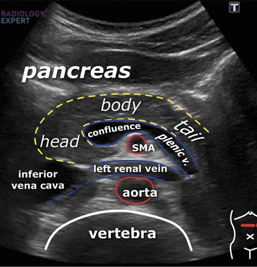
pancreatitis
appendicitis
testicular problems
FAQ
How does an ultrasound scan work?
An ultrasound scan works by using high-frequency sound waves to create images of parts inside the body. These sound waves are emitted from a small probe called a transducer and are reflected to internal organs, tissues, and fluids. The reflected waves are then captured by the transducer and converted into images that can be viewed on a monitor.
What are the advantages of an ultrasound scan over other imaging techniques?
Ultrasound scans offer several advantages over other imaging techniques, like MRI or CT scans. They are non-invasive, painless, and do not involve radiation exposure, making them safer for pregnant women and children. Ultrasound scans can also provide real-time images, allowing for immediate evaluation and diagnosis. Additionally, they are cost-effective than other imaging scans and can be used to guide procedures such as biopsies or injections.
Are there any risks associated with ultrasound scan tests?
Ultrasound scan tests are generally considered safe, with no known risks or harmful effects associated with the procedure. However, excessive or unnecessary use of ultrasound should be avoided.
How long does an ultrasound scan take to progress?
The ultrasound scan will usually take 20 to 30 minutes to complete. However, scanning of vascular parts like arteries and vessels, where the organs are increased in number and size, may take longer.